Science
Understanding Degeneration
Cajal Neuroscience is a drug discovery company built for one purpose: to deliver new medicines that give life back to patients suffering from neurodegenerative disease.
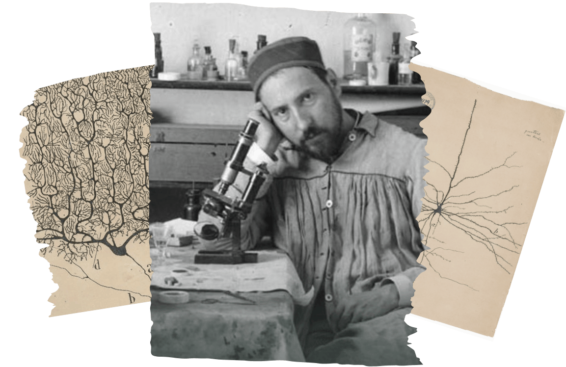

We are inspired by the pioneering work of Santiago Ramón y Cajal who in the late 1800s illustrated the cellular content and connectivity of the brain in remarkable detail.
From his intense devotion to visualizing the brain at cellular resolution, Cajal´s discoveries on the structural and functional organization of the brain have since become the foundation of modern neuroscience. Similarly, to truly understand the complex mechanisms of neurodegeneration and how we can intervene, we must be able to better visualize it.
Our approach is to build a platform for target discovery and validation, one of unprecedented scope and depth that not only allows us to “see” what is happening in the degenerating brain, but also where and when. Accomplishing this feat requires a unique marriage of human genetics and multi-omics techniques, next-generation sequencing and imaging technologies, and both cellular and animal models of disease.
Drivers of Neurodegeneration
Advances in genetics have transformed our understanding of neurodegeneration, yielding expansive lists of genes and pathways associated with disease. However, translating this wealth of data into therapeutic insight remains challenging and requires understanding how, where, and when these genes and pathways mediate disease risk and progression.
At Cajal, we apply powerful multi-omics methods and multiplexed in vitro and in vivo screens to systematically prioritize and functionally validate the thousands of genes implicated in neurodegenerative diseases. Through this approach, we aim to identify novel targets most likely to modify the course of neurodegeneration.
Changing the Course of Neurodegeneration
A fundamental challenge in neurodegeneration has been the inability to access human molecular data during disease. Genetics teaches us about the pathways contributing to disease onset, while post-mortem pathology depicts the aftermath of disease. Understanding what is happening during the actual course of neurodegeneration, and how to change it, requires new approaches to modeling and visualizing disease processes.
We combine deep expertise in translational models with state-of-the-art tools, from lightsheet microscopy to high content imaging, to interrogate novel therapeutic hypotheses with greater spatiotemporal precision. The flexibility and scalability of our assays allow us to evaluate the function of numerous targets in parallel, or drill deep into the unique mechanistic biology of a specific target or drug candidate.
Ref. Lightsheet microscopy-generated 3D visualization of mouse brain showing amyloid beta plaques (blue) and axonal projections (orange)
Visualizing Neurodegeneration
Our industrialized lightsheet microscopy platform allows us to directly visualize the biological effects of a target or therapeutic in the intact brain. With 3D imaging, we can evaluate complex spatial phenotypes like selective vulnerability or spread of pathology.
We leverage multiple other methods in parallel, such as sequencing-based axon mapping, scRNAseq, and a range of in vitro assays, to create a holistic image of target or drug function.